


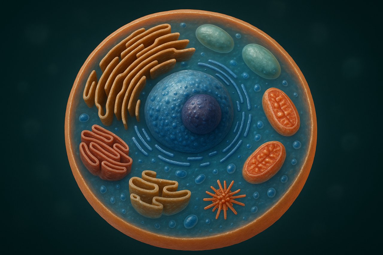
Understanding the animal cell diagram is fundamental to mastering biology. Every organism from a microscopic protozoan to complex mammals depends on the functions of cells. The animal cell is the basic structural and functional unit of all animal life. In this article, we’ll explore the complete animal cell diagram, identifying each organelle, explaining its function, and connecting it to biological processes that sustain life.
Examine the Overall Structure of the Animal Cell Diagram

An animal cell is a eukaryotic cell, meaning it has a well-defined nucleus enclosed by a nuclear membrane and numerous membrane-bound organelles. The diagram of an animal cell usually shows a spherical or irregular shape, enclosed by a cell membrane that separates the internal environment (cytoplasm) from the external environment.
At the center of the diagram lies the nucleus, surrounded by various organelles like the mitochondria, endoplasmic reticulum, Golgi apparatus, and lysosomes. Each organelle performs specialized functions, but together they maintain homeostasis, enabling the cell to survive, grow, and reproduce.
The animal cell diagram also reveals that unlike plant cells, animal cells lack a cell wall and chloroplasts, which gives them flexibility and mobility. Their irregular shape allows animal tissues to adapt to diverse physiological roles muscle contraction, nerve impulse transmission, and immune defense.
Shape and Boundary of Animal Cells
Animal cells lack a rigid cell wall, giving them flexibility and irregular forms. This flexibility allows for movement and complex tissue formation something not possible in plant cells. The outermost boundary is the plasma membrane, which regulates what enters and leaves the cell, maintaining an internal balance known as homeostasis.
Key Features That Define Eukaryotic Cells
Inside the boundary, the cytoplasm contains various membrane-bound organelles tiny, specialized structures performing distinct functions. Central to this system is the nucleus, the command center that houses DNA. Together, these organelles form a coordinated system that enables the cell to grow, repair, and reproduce efficiently.
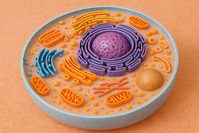
Identify the Key Organelles in the Animal Cell Diagram
Every labeled animal cell diagram features major organelles that represent unique biological roles. These structures include the nucleus, mitochondria, ribosomes, endoplasmic reticulum (ER), Golgi apparatus, lysosomes, peroxisomes, and centrosomes.
Major Membrane-Bound Organelles
Membrane-bound organelles include the nucleus, ER, Golgi apparatus, mitochondria, lysosomes, and peroxisomes. These organelles have their own compartments that separate biochemical reactions, ensuring efficiency and control.
Non-Membrane Organelles and Their Roles
Ribosomes and centrosomes are non-membranous but critical for protein synthesis and cell division, respectively. Their presence ensures cellular maintenance and reproduction.
| Organelle | Structure | Primary Function |
| Nucleus | Double membrane enclosing DNA | Controls genetic activity |
| Mitochondria | Double membrane, inner folds (cristae) | Produces ATP through respiration |
| Ribosomes | RNA-protein complexes | Protein synthesis |
| ER (Rough/Smooth) | Interconnected membrane network | Synthesizes proteins/lipids |
| Golgi Apparatus | Flattened stacked membranes | Modifies and packages proteins |
| Lysosomes | Enzyme-filled vesicles | Digests waste |
| Peroxisomes | Small oxidase-containing vesicles | Detoxifies harmful substances |
| Centrosome | Two centrioles | Aids in cell division |
Understand the Role of the Nucleus and Nucleolus
The nucleus is often labeled as the control center of the cell because it regulates genetic expression and cellular reproduction. In the diagram, it appears as a large circular structure with a double-layered nuclear membrane containing pores. These pores act as gateways, allowing the movement of mRNA, ribosomal subunits, and enzymes between the nucleus and cytoplasm.
Inside the nucleus, the nucleolus is a darkly stained spherical body responsible for producing ribosomal RNA (rRNA) and assembling ribosome subunits. This nucleus-nucleolus complex governs protein synthesis by sending instructions in the form of messenger RNA (mRNA) to ribosomes.
From a biological perspective, the nucleus acts as the information repository, while the nucleolus functions as the machinery initiator. In animal cell diagrams, both are drawn prominently because they define the cell’s identity and heredity.
Nuclear Envelope and Genetic Material
The nucleus is enclosed by a double-layered nuclear membrane containing nuclear pores that regulate molecular traffic between the nucleus and cytoplasm. Inside, chromatin (DNA and proteins) holds the genetic blueprint of life. During cell division, chromatin condenses into chromosomes that carry genetic information to new cells.
Function of the Nucleolus in Ribosome Formation
Within the nucleus lies the nucleolus, a dense region responsible for producing ribosomal RNA (rRNA). It assembles ribosome subunits that later move into the cytoplasm to initiate protein synthesis. The cooperation between the nucleus and nucleolus ensures the cell produces necessary enzymes and structural proteins for survival.
Analyze the Cytoplasm and Its Supporting Matrix
The cytoplasm is the semi-fluid matrix that fills the cell, supporting organelles and facilitating the transport of materials. It is composed of cytosol (a gel-like substance) and a cytoskeleton (a network of fibers such as microtubules, actin filaments, and intermediate filaments).
The cytoskeleton provides structural integrity and enables intracellular movement. Microtubules act as tracks for motor proteins, while actin filaments are crucial for cell shape and movement. Intermediate filaments provide mechanical support, particularly in cells subjected to stress like epithelial cells.
In the animal cell diagram, the cytoplasm appears as the light-colored background where all organelles are embedded. Despite being often overlooked, it’s a dynamic component that influences everything from vesicle transport to cell signaling.
Cytosol and Chemical Reactions
The cytosol, which forms the fluid portion of the cytoplasm, contains ions, enzymes, and dissolved nutrients. It serves as the site for countless metabolic reactions such as glycolysis, maintaining energy flow within the cell.
Cytoskeleton and Structural Support
The cytoskeleton, made up of microtubules, actin filaments, and intermediate filaments, provides mechanical support and movement. It also acts as a transport network for vesicles and organelles. This internal scaffolding system is vital for maintaining cell shape and enabling motility in animal cells.
Examine the Function of the Cell Membrane
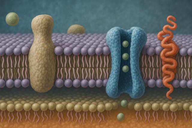
The cell membrane, also known as the plasma membrane, forms the outer boundary of the animal cell. It is composed of a phospholipid bilayer with embedded proteins, cholesterol molecules, and carbohydrates.
This membrane performs selective permeability, allowing only certain molecules (like oxygen, nutrients, and ions) to enter or leave the cell. Embedded proteins act as transporters, receptors, and enzymes, controlling communication and material exchange.
The flexibility of the plasma membrane, as seen in the diagram, allows animal cells to change shape, engulf particles (endocytosis), and release substances (exocytosis). This dynamic nature supports cell signaling, nutrient uptake, and immune recognition.
Phospholipid Bilayer Structure
The membrane’s structure consists of a phospholipid bilayer with embedded proteins, cholesterol, and carbohydrates. The hydrophilic (water-attracting) heads and hydrophobic (water-repelling) tails create a semipermeable barrier that maintains chemical balance.
Transport and Communication
Transport proteins regulate the flow of substances like ions and nutrients, while receptor proteins facilitate communication through signal transduction. This structure allows the animal cell to perform processes like endocytosis (intake of materials) and exocytosis (release of materials).
Study the Mitochondria as the Powerhouse of the Cell
The mitochondria are shown as oval-shaped organelles with inner folds called cristae. These cristae increase surface area for biochemical reactions that generate adenosine triphosphate (ATP) the cell’s energy currency.
Through the process of cellular respiration, mitochondria convert glucose and oxygen into energy (ATP), water, and carbon dioxide. The inner mitochondrial matrix contains enzymes for the Krebs cycle, while the inner membrane hosts the electron transport chain.
Interestingly, mitochondria have their own DNA and ribosomes, suggesting an evolutionary origin from free-living bacteria (endosymbiotic theory). In the animal cell diagram, mitochondria are usually depicted throughout the cytoplasm, representing their high distribution in energy-demanding cells like muscles and neurons.
Structure and Inner Cristae
The mitochondrion has a double membrane an outer membrane and an inner membrane with folds called cristae. These folds increase surface area, hosting enzymes crucial for ATP synthesis.
ATP Production Process
Through cellular respiration, mitochondria break down glucose in the presence of oxygen to produce ATP, water, and carbon dioxide. Because mitochondria contain their own DNA and ribosomes, they can replicate independently, hinting at their evolutionary origin as free-living bacteria.
Explore the Endoplasmic Reticulum (ER) Network
The endoplasmic reticulum (ER) is a complex network of membranes continuous with the nuclear envelope. It comes in two forms: rough ER (RER) and smooth ER (SER).
The Rough ER is covered with ribosomes and is responsible for protein synthesis. Newly synthesized proteins enter its lumen for folding and modification. The Smooth ER, lacking ribosomes, performs lipid synthesis, detoxification, and calcium storage.
In the animal cell diagram, RER is drawn close to the nucleus, reflecting its role in genetic expression and protein production. SER is distributed more peripherally, emphasizing metabolic and detoxification processes.
Rough Endoplasmic Reticulum (RER)
RER has ribosomes attached to its surface, giving it a “rough” appearance. It’s primarily involved in protein synthesis and modification. Once synthesized, proteins are transported to the Golgi apparatus for further processing.
Smooth Endoplasmic Reticulum (SER)
The SER lacks ribosomes and is responsible for lipid synthesis, carbohydrate metabolism, and detoxification. In muscle cells, it also regulates calcium ion levels necessary for contraction.
Understand the Golgi Apparatus and Protein Packaging
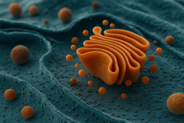
The Golgi apparatus (or Golgi complex) is shown as a series of flattened, stacked membranes called cisternae. It functions as the processing and packaging center of the cell.
Proteins and lipids produced by the ER are transported in vesicles to the Golgi apparatus. There, they are modified glycosylated, folded, or sorted and then packed into new vesicles destined for secretion or lysosomal storage.
In the animal cell diagram, the Golgi apparatus is often located near the nucleus and ER, indicating close cooperation in the protein synthesis and secretion pathway. Its role ensures the proper distribution of proteins within the cell and outside it, a process crucial in hormone secretion and immune responses.
Cis and Trans Faces of Golgi Apparatus
The Golgi has two faces the cis face, which receives vesicles from the ER, and the trans face, which ships modified products to their destinations.
Modification and Secretion
Proteins and lipids are modified, sorted, and packed into vesicles. Some are sent to the plasma membrane for secretion, while others are transported to lysosomes. This step is vital for hormonal and enzymatic secretion in animal systems.
Detail the Lysosomes and Cellular Digestion
Lysosomes are small, spherical organelles filled with digestive enzymes. Their primary function is to break down waste materials, defective organelles, and foreign particles.
These enzymes function optimally in acidic conditions. When a cell engulfs pathogens or debris, lysosomes fuse with the vesicle and digest the contents a process known as autophagy or phagocytosis.
In the diagram, lysosomes are depicted as small circular bodies scattered through the cytoplasm. Their activity is essential for cellular renewal, defense, and programmed cell death (apoptosis).
Structure and Enzymes
Each lysosome is surrounded by a membrane that maintains an acidic internal environment, optimal for enzymatic activity. The enzymes include proteases, lipases, and nucleases that degrade biological molecules.
Autophagy and Defense
Lysosomes perform autophagy (digestion of damaged organelles) and phagocytosis (digestion of external particles). This process keeps the cell clean and protects against infection.
Investigate the Role of Peroxisomes and Centrosomes
Peroxisomes resemble lysosomes but have different enzymes, primarily oxidases and catalases, which detoxify substances like hydrogen peroxide. They are vital in fat metabolism and detoxification processes within liver cells.
Centrosomes consist of two centrioles positioned at right angles near the nucleus. They play a key role in cell division, organizing microtubules into spindle fibers that separate chromosomes during mitosis.
In the animal cell diagram, centrosomes are often labeled near the nucleus, while peroxisomes appear dispersed. Together, they illustrate how cells maintain order during division and detoxify themselves simultaneously.
Peroxisomes in Detoxification
Peroxisomes contain oxidative enzymes that detoxify harmful compounds, especially in liver and kidney cells. They convert toxic hydrogen peroxide into water and oxygen using the enzyme catalase.
Centrosomes in Cell Division
The centrosome, composed of two centrioles, organizes microtubules during mitosis. It helps form the spindle apparatus, ensuring proper chromosome segregation.
Differentiate Between Animal and Plant Cell Diagrams
Although both plant and animal cells are eukaryotic, their diagrams differ significantly due to structural and functional variations.
| Feature | Animal Cell | Plant Cell |
| Cell Wall | Absent | Present (made of cellulose) |
| Chloroplasts | Absent | Present for photosynthesis |
| Shape | Irregular, flexible | Rectangular or rigid |
| Vacuole | Small and temporary | Large central vacuole |
| Energy Source | Mitochondria | Mitochondria + Chloroplasts |
| Lysosomes | Common | Rare or absent |
Structural Differences
Animal cells lack a cell wall and chloroplasts, whereas plant cells possess both. This makes animal cells more flexible, while plant cells maintain rigidity.
Functional and Metabolic Variations
Plant cells produce food via photosynthesis using chloroplasts, while animal cells rely on cellular respiration to convert nutrients into energy.
| Feature | Animal Cell | Plant Cell |
| Cell Wall | Absent | Present |
| Chloroplasts | Absent | Present |
| Vacuole | Small, temporary | Large central vacuole |
| Shape | Irregular | Rectangular |
| Energy Source | Mitochondria | Mitochondria + Chloroplasts |
Illustrate Cell Division through the Animal Cell Diagram
Cell division is represented in the diagram by the changing shape of the nucleus and centrosomes. Animal cells divide through mitosis (for growth and repair) and meiosis (for reproduction).
During mitosis, the centrosomes form spindle fibers that pull apart replicated chromosomes into two daughter cells. Each new cell inherits a complete set of genetic material.
In meiosis, genetic material is reduced by half, forming gametes (sperm and eggs). The diagram of dividing animal cells shows chromatin condensation, nuclear membrane breakdown, and chromosome segregation steps that ensure genetic continuity.
Mitosis and Its Phases
Animal cells divide through mitosis, producing two genetically identical daughter cells. The process includes prophase, metaphase, anaphase, and telophase, followed by cytokinesis.
Meiosis and Genetic Diversity
In reproductive cells, meiosis produces gametes with half the chromosome number, introducing genetic diversity. The centrosomes and spindle fibers illustrated in the animal cell diagram are critical for these processes.
Interpret the Importance of Vesicular Transport
Within the cytoplasm, the endomembrane system operates as a coordinated network of vesicles transporting materials. Vesicular transport connects the ER, Golgi apparatus, plasma membrane, and lysosomes.
There are two main types:
- Endocytosis (intake of materials)
- Exocytosis (release of materials)
In an animal cell diagram, vesicles are often depicted as small bubbles moving between organelles. This highlights the dynamic flow of biomolecules necessary for cell communication, metabolism, and waste removal.
Endocytosis and Exocytosis
Vesicular transport involves endocytosis (intake of substances) and exocytosis (release of substances). These processes move molecules like proteins and lipids between the ER, Golgi, and plasma membrane.
Relate the Animal Cell Diagram to Real-World Applications
Understanding the animal cell structure has profound applications in medicine, genetics, and biotechnology.
- In medicine, cellular knowledge enables tissue engineering, cancer research, and regenerative therapies.
- In genetics, it aids in gene editing (CRISPR), DNA replication studies, and hereditary disease mapping.
Therefore, mastering the animal cell diagram is not just academic; it’s the foundation for innovations that sustain life sciences and modern healthcare.
Medical and Genetic Research
Knowledge of the animal cell diagram supports cancer research, gene therapy, and drug development. Understanding how organelles function helps identify cellular abnormalities.
Conclusion:
The animal cell diagram serves as a visual blueprint of life’s complexity. Every organelle from the nucleus to the mitochondria works in harmony to sustain cellular processes. Understanding these structures deepens comprehension of human physiology, medical science, and biotechnology.
Cells are not mere components; they are dynamic factories of life. The more we understand their diagrams and mechanisms, the closer we come to understanding how all living systems operate and evolve.
Explore more insightful and valuable content on our blog VeoTag.com! Stay updated with helpful tips, expert advice, and in-depth articles that enhance your knowledge.
Read Also:
1. kiddle: The Safe Search Engine That Makes the Web Kid-Friendly
2. Chase Cominsky: Legal Cases & Fallout of the Lake Erie Fishing Cheating
3. Halloween Jokes: Funny, Family-Friendly & Spooky Puns for 2025
4. istreameast: Is It Safe, How It Works & Legal Alternatives
5. Salty Ice Cream: Savory Frozen Desserts Are the Next Big Trend
6. Pumpkin Painting: Step-by-Step Guide for Stunning Decor
7. CrackStreams: Legal Risks & Best Alternatives in 2025
8. simpcity: Inside the Digital Culture of Fandom, Leaks & Community
FAQ’s
It visually represents the structure and functions of animal cells, helping students and scientists understand cellular organization.
Because animals rely on ingestion for energy, not photosynthesis. The absence of a rigid cell wall gives animal cells flexibility.
The cell membrane, cytoplasm, and nucleus are the three core components of an animal cell.
The mitochondrion generates ATP, providing energy for all cellular functions.
It modifies, sorts, and packages proteins and lipids for secretion or use within the cell.
They digest waste, destroy pathogens, and recycle damaged organelles to maintain cell health.


